When artist Leonor Caraballo was diagnosed with breast cancer in 2008, the small lump in her left breast was described by her doctor as “aggressive but small.” The shape of the thing wasn’t made clear, just that it was 1.3 centimeters. “It’s kind of abstract,” she said. “I had this clichéd notion that it looked like a golf ball.”
For many like Caraballo, along with all the terror and dread of diagnosis, there comes a mental image of a dark, ominous orb gnawing its way through flesh. And as Caraballo and her husband and artistic partner Abou Farman were about to learn, that picture was all wrong. Ahead would be confirmation that Caraballo is a BRCA2 carrier, with heightened breast cancer risk. There would be a mastectomy in 2008, an oophorectomy in early 2009. But over the course of medical treatment, Farman remembers Caraballo saying, “I need to know what this looks like, because it’s in me. I still have no idea what it is.”
“I didn’t like not knowing how big it was and what shape it had and what form it had. It bothered me,” Caraballo said.
Perhaps it is natural that an artist would want to properly visualize her tumor. The couple sat with radiologists and asked to see what they saw. They learned about the software the medical team used, how they manipulated MRIs. Farman remembers a shared sense that “these are incredible technologies. They are showing incredible things inside, and it’s useful to see them. And it’s comforting also to be able to sit there and see this thing—and get the measure of it, in a sense.”
Just months after Caraballo completed surgery, the multimedia artists, working together under the moniker caraballo-farman, began a six-month process of trial and error, trying to figure out ways to blueprint tumors three-dimensionally, using MRIs from cancer patients and friends, with an eventual goal of using a 3-D printer to replicate them. They had a variety of tools—fellowships from the Guggenheim Foundation and the New York Foundation for the Arts, a residency at New York’s art and technology center Eyebeam, and help from doctors at Weill Cornell Medical Center and NYU Langone Medical Center. But each step was a challenge because they were navigating uncharted territory—both artistically and medically—in learning how to manipulate the MRIs to create replicas you could hold in the palm of your hand. “Every time we managed to isolate something from a tumor, it was like, Yes! One point of triumph, then the next step.” Finally, continues Farman, “we cleaned it up and hit print, and this thing came out.”
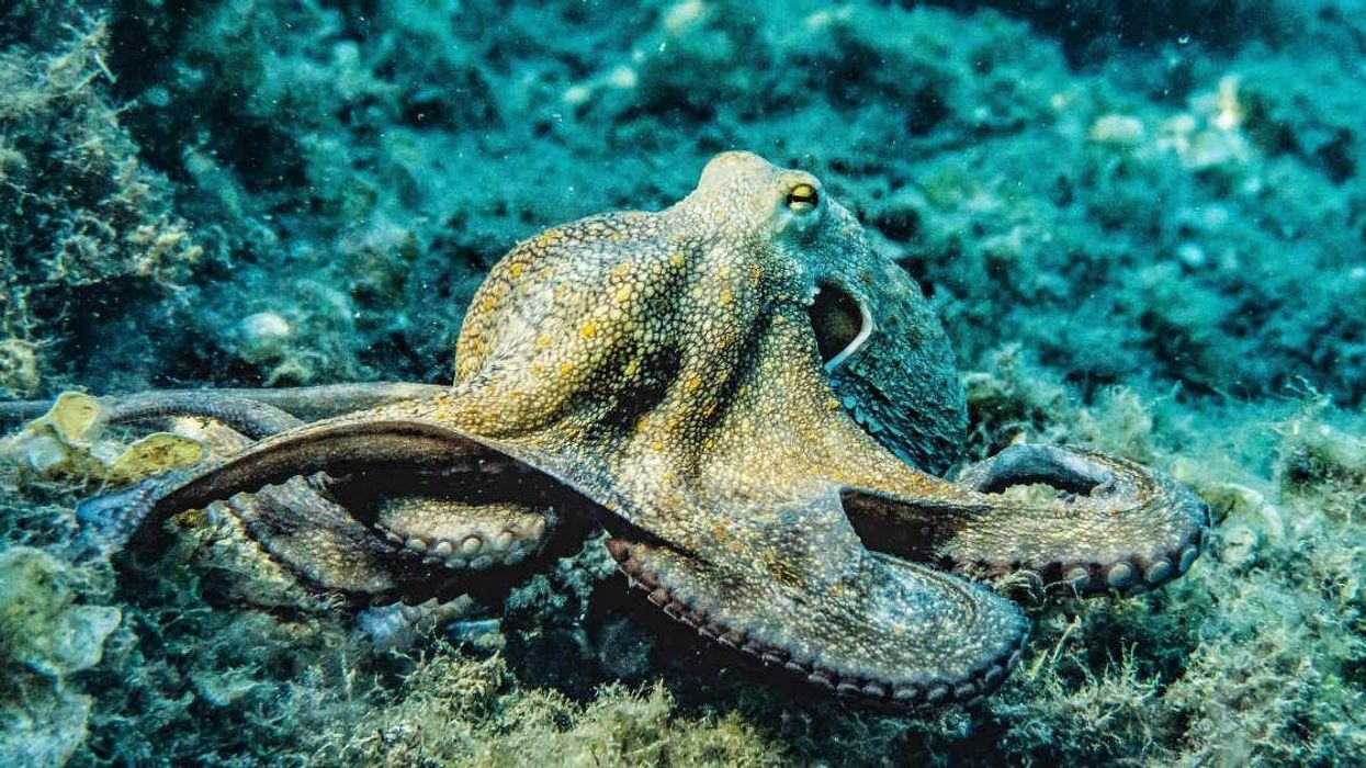
The result was less a shapeless orb than something akin to a chunk of coral. “They look organic,” said Caraballo, “like sea creatures” with spiculations “like tentacles”—that grow to invade new areas. First there were 3-D prints in ABS plastic, then casts and large-scale bronze pieces, performance pieces, prints. “By externalizing it,” said Caraballo, “I made it a solid. I made it into a rock that couldn’t move or change anymore, metaphorically.”
The tumors became two different types of art. One, small, able to be carried or worn. For a disease that is still often voiced in whispers, cancer could become a conversation piece, a tumor, an invisible thing turned visible and able to be manipulated by a patient. It would also be a way to cut through the remaining taboo surrounding cancer. “I think part of the phobia is that it’s not spoken of enough, or it’s spoken of with fear and trepidation and not in a direct way,” said Farman.
Caraballo explains that they wanted to create a worry bead, an amulet, something to ward off evil spirits. Alternatively, said Caraballo, “it wouldn’t necessarily have to be regarded as anything precious… I almost wanted to give it to the dog to chew on or whatever, throw it in the river, bury it, do a ritual around it. To make it alive outside the body.” Charms, pendants, paper weights and worry beads, in the shape of tumors, can be purchased from the Object Breast Cancer website.
And seeing cancer anew—even for those who spend their days treating and tending to cancer patients—has been a radical shift. Caraballo-farman’s first show of large form tumor sculptures was attended by a number of oncologists and surgeons. Says Farman, “They were kind of struck by something that they hadn’t seen, because although they look at it on a screen, they had never seen it as a full-on object that they could walk around. And it struck them that it should become, not just an art project, but a medical project.”
Caraballo’s doctor, Dr. Alexander Swistel, breast cancer surgeon at New York-Presbyterian Hospital/Weill Cornell Medical Center, explains that caraballo-farman’s work “absolutely inspired me to re-think how we stage breast cancers.” Generally, Swistel explains, staging is based on uni-dimensional measurements, but the caraballo-farman demonstration of three-dimensional tumor growth “showed me that volume measurement may be more pertinent.”
Based on that observation, and in collaboration with Weill Cornell radiologist Michelle Drotman, the doctor has started a retrospective review to see if traditionally staging malignancies uni-dimensionally resulted in over- or under-treatment, as compared to what would have been prescribed using volumetric, three-dimensional measurements. They are also investigating through clinical trials to see if volumetric measurements could affect oncologists' chemotherapy recommendations. Swistel notes that results are still pending, but “I have no doubt that this may in fact revolutionize the way we think about staging cancer of the breast and other solid tumors as well.”
The desire to see the tumor in its true form initially, says Farman, “wasn’t strictly an artistic impulse, but it came out of an artistic place.” And now art, having imitated disease, could quite possibly have inspired a better way to save lives.







 Otis knew before they did.