Nature’s sporting a new look that’s straight out of a science fiction movie. Scientists in China were able to genetically engineer succulents that glow in the dark. There have been other recent attempts at implementing the creative new technology, but they were limited to only a green color. Now, there are some wild variations, and the plants might one day create the perfect lighting for a futuristic rave.
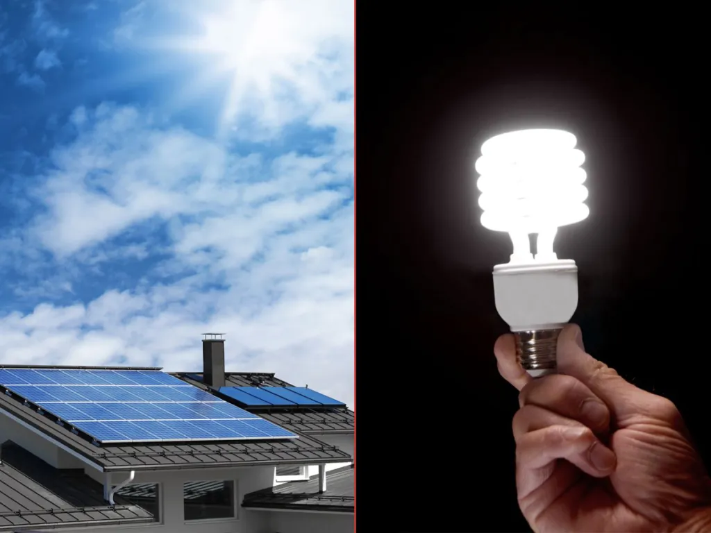
This science, over time, could lead to many variations of lighting that could possibly replace the need for electricity. These glow-in-the-dark succulents recharge with sunlight, and the colorful displays are absolutely magical.
Translucent succulents in the dark. Image credit Liu et al., Matter
First author Shuting Liu of South China Agricultural University was quoted in a 2025 article of EurekaAlert! saying, “Picture the world of Avatar, where glowing plants light up [the] entire ecosystem.” She continues, “We wanted to make that vision possible using material we already work with in the lab. Imagine glowing trees replacing streetlights.”
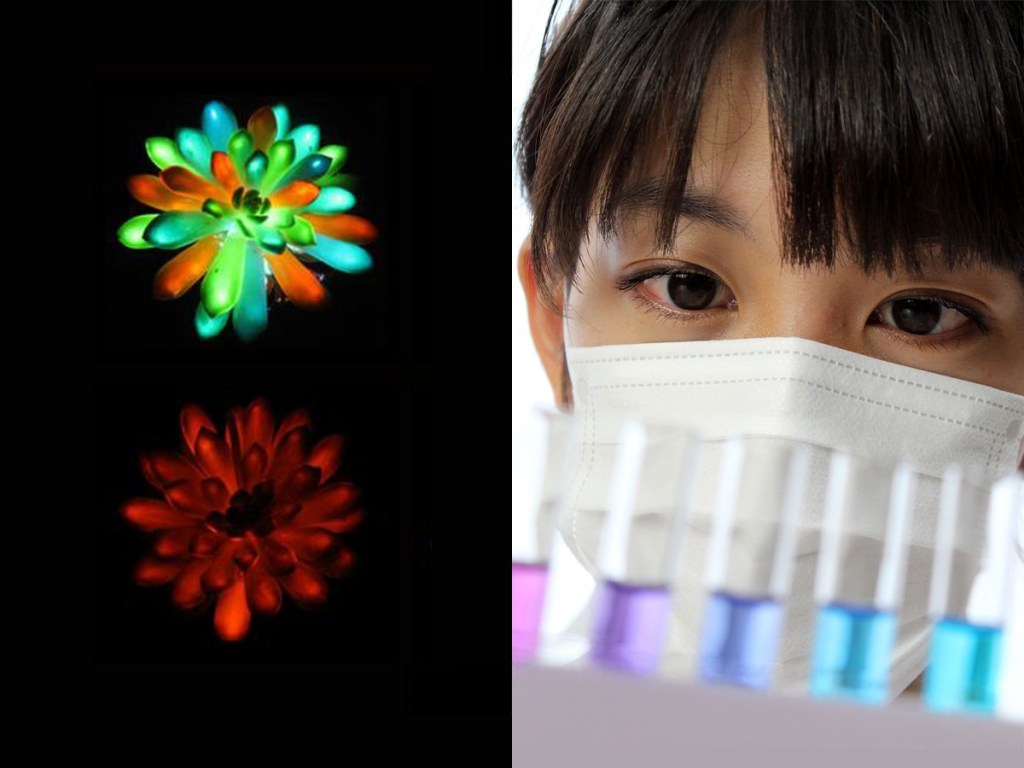
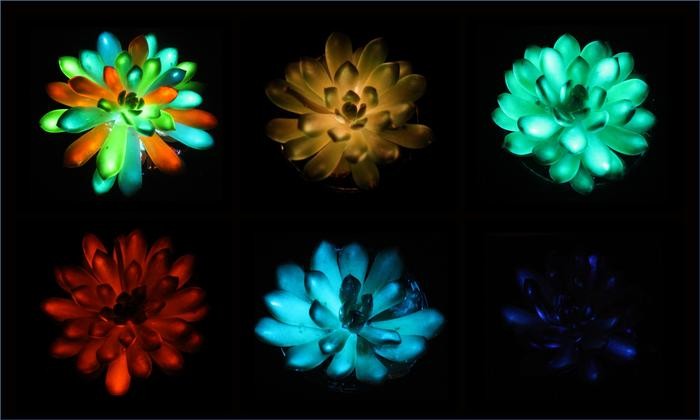
Using different kinds of phosphors, they were able to create plants that shine in various colors.
[html5_video https://roar-assets-auto.rbl.ms/runner%2Fmmc4.mp4 url=”https://roar-assets-auto.rbl.ms/runner%2Fmmc4.mp4″ shortcode_id=1756482759483 videoControls=true feedbacks=true mime_type=”video/mp4″ expand=1 site_id=26881454 caption=”Injecting phosphors into a succulent.” photo_credit_src=”https://roar-assets-auto.rbl.ms/runner%2Fmmc4.mp4″ photo_credit=”Vidoe credit Liu et al., Matteru00a0 u00a0roar-assets-auto.rbl.ms”]
The study appeared in a 2025 article of Matter, a sister journal to Cell Press, stating, “Plant-based lighting holds significant potential across various fields, including architecture and urban planning. However, manipulating luminescence color and intensity in plants has been challenging. Traditional genetic engineering approaches are constrained by the limited diversity of bioluminescent genes.” Using light-emitting phosphor particles that were about the size of a human red blood cell (around seven micrometers), they were able to produce a glow that would travel through the biological structure of the succulent plants. “Smaller, nano-sized particles move easily within the plant but are dimmer,” Liu claimed. “Larger particles glowed brighter but couldn’t travel far inside the plant.”
To achieve stronger visible luminescence, the light-emitting particles had to be large enough to emit the glow yet small enough to travel through the plant’s tissue. Other plants were able to absorb the phosphorus, but the composition of the plant tissues wasn’t effective for emitting a significant and well-distributed glow. Succulents appear to be fairly successful. “I just find it incredible that an entirely human-made, micro-scale material can come together so seamlessly with the natural structure of a plant,” says Liu. “The way they integrate is almost magical. It creates a special kind of functionality.”
The process takes about 10 minutes to prepare a plant, and the cost, including labor, is less than $2, according to Liu. “It was really unexpected…the particles diffused in just seconds, and the entire succulent leaf glowed.”
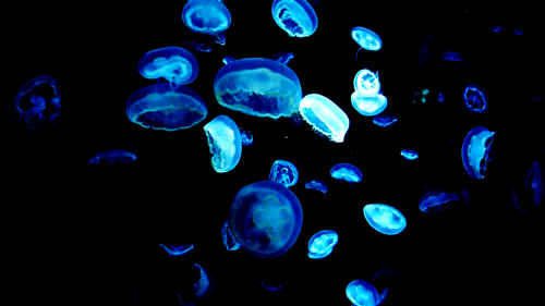
A 2025 study published in the British Ecological Society found that sheet web spiders would trap and use fireflies to lure insects into their webs. Having the bioluminescent fireflies attracted significantly more prey. Getting crafty isn’t just left to the spiders, though. Other forms of nature using this biotech might be more familiar. Images of glowing jellyfish have long been at the forefront of nature books and science magazines. However, scientists believe the beginnings of bioluminescence started with deep-sea coral. According to a 2024 study reported by the Associated Press, researchers analyzing genetic data from 185 species of glowing coral believe their ancestors first lived and “glowed” about 540 million years ago.
It’s fantastic what marvels science and ingenuity can bring to the normal lives of everyday people. By combining natural structures, such as plants, with man-made materials, science may open the door to sustainable, plant-based lighting for future cities. This breakthrough demonstrates how engineered light-emitting particles can seamlessly integrate with plants. With new and affordable ways to make plants glow, the future looks bright indeed.
Plastic waste has been a growing global issue for years. The United Nations Environment Programme says that 19 to 23 million metric tonnes of plastic waste leaks into lakes, rivers, and oceans each year. Given the threat microplastics pose to animal and human health, efforts to find green replacements have intensified—and they’re working. Scientists from Singapore and Spain have found a strong, potentially viable replacement for plastic made from shrimp shells.
A research team based at the Singapore University of Technology and Design and the Institute for Bioengineering of Catalonia in Barcelona has made a biodegradable plastic alternative out of chitosan. Chitosan is a compound created by combining shrimp shells with trace amounts of nickel. It contains a structural molecule found in the shells of crustaceans and insect exoskeletons. Usually discarded as a waste byproduct of shrimp and crab processing, chitosan is commonly produced during seafood preparation and commercial fishing.
The issue with chitosan, though, was that it weakens and dissolves in water. That is, until recently.
How chitosan got stronger
Dissolving chitosan flakes into a weak acetic solution and mixing them with dissolved nickel chloride and water produced surprising results. Scientists then poured the mixture into molds to dry. The process yields a thin, green-tinted film with the strength of commonly used plastics like polypropylene. Even better, when submerged in water, the film grows 50% stronger. This increased durability matches the characteristics of polycarbonate and PETG, plastics commonly used in commercial single-use water bottles.
Researchers then stress-tested the chitosan material by molding it into cups and containers. They were able to confirm it could hold water without leaks. In terms of biodegradability, the chitosan material reached its half-life in four months in a standard soil burial test. By contrast, most commercial plastics can take centuries to decompose under similar conditions.
Researchers found that this is not only a better biodegradable plastic alternative, but also one that produces zero waste during creation. When the chitosan/nickel film is submerged, about 87% of the nickel washes out. That wash water can then be reused again and again from one batch of chitosan to the next. According to the researchers, the nickel content of a single AAA battery would provide enough nickel to manufacture more than a dozen chitosan drinking cups.
The potential future
Rigorous testing to assess the material’s limits for medical use and consumption still needs to be done. That said, the Food and Drug Administration has already approved products containing chitosan and nickel individually in the past. Barring any troubling research about their combined safety, the outlook is quite positive for future use.
Hopefully, seafood and battery waste can be reduced, helping lower plastic waste in a three-way win for the environment.
In the United States, gut health is a big deal. According to a 2022 survey by the American Gastroenterological Association, 40% of Americans deal with digestive problems that disrupt their day-to-day lives. Many try different diets and supplements to assuage these issues, as well as use probiotics to improve overall digestion—but there might be a better way. Scientists may have found a way to analyze and pinpoint potential digestive problems; all you need to do is put on some special underwear and fart.
Researchers at the University of Maryland wanted to find a better way to monitor and measure human gut bacteria in the name of microbiome research. While past methods allowed them to see what gut bacteria species are living in the human body, there was no accurate way to see what the gut bacteria was doing hour by hour. Well, they seem to have found a way: a pair of underwear with a tiny sensor clipped near the rear can now record data from a person’s flatulence.
This “Smart Underwear” measures the amount of hydrogen gas emitted each time a person passes gas, monitoring the gut bacteria’s activity within a person. Hydrogen gas is typically produced when certain gut bacteria breaks down undigested food. The whole thing may sound silly (and smelly), but early tests of this device have been able to detect dietary changes in people with 94.7% accuracy. This device and method of analyzing gut bacteria is better than most current tools that analyze stool samples, blood, or breath for such data. The Smart Underwear also has a battery that can last for a week without compromising comfort for the wearer.
“The Smart Underwear comfortably attaches to the exterior of the user’s underwear near the perineal region via a snap system, in which a small plastic snap on the inside of the underwear fabric fits into a corresponding hole on the Smart Underwear on the opposite side of the underwear,” wrote Santiago Botasini and their colleagues in the study. “This sandwiches the fabric in place through friction, ensuring a stable but comfortable attachment of the Smart Underwear. Once attached, the Smart Underwear passively captures hydrogen concentration in flatus, as well as temporal dynamics including the frequency and duration of flatus events enabling longitudinal measurements of gut microbial metabolism.”
“Healthy adults produced flatus an average of 32 times per day” #SmartUnderwear#Flatus is the medical term for gas that is expelled from the digestive tract through the rectum #Fartshttps://t.co/9OO7XVbmXW
While additional studies are needed, this current study of 38 participants seems to suggest this device could help doctors. The Smart Underwear could pinpoint specific food sensitivities and intolerances within their patients, which proves to be a much better and more accurate practice than relying on patients self-reporting via “food journals” to capture patterns.
The study shows that self-reporting isn’t always accurate. For example, the device revealed that the participants farted 32 times per day on average, which is more than double the typically cited daily average of 14 incidents.
It will be a while before gastrointestinal doctors start prescribing Smart Underwear to figure out what’s causing their digestive discomfort, but the research is promising. At any rate, whether the Smart Underwear will be used just for studies or becomes a widespread method to identify food intolerances, getting more information will ultimately lead to a sweeter smell of success over time.
Today the world of Egyptology faces a silent crisis – not of looting, although that plays a part, but of disconnection. Walk into any major museum, from Copenhagen to California, and you see glass cases filled with what could be called orphaned artifacts: remarkable objects, often acquired in the 19th and early 20th century, that have been completely stripped of their histories. You can see what they are – a mummy’s painted foot case, a golden mask – but we have no idea where they came from. They are beautiful, but historically they are mute.
Many objects entered museum collections at times when excavation and collecting practices were very different from today. In the past, excavated objects were often divided between institutions around the world, and display was prioritized over documentation. Over time, connections between pieces were lost. As a result, museums around the world hold remarkable artifacts whose backstories are thin, fragmentary or missing altogether.
Archaeologists like me working in the field today regularly uncover fragments: broken pieces of objects that once formed part of something larger. In some cases, those fragments may share the same underlying geometry with objects already held in museums. For example, a mummy’s foot case and a newly found shard may have been produced using the same mold, so they share a consistent three-dimensional form even if they are now separated by time, distance and absence of documentation.
Traditionally, evaluating whether a fragment matches up with a specific museum object has relied on visual judgment and incomplete records, rather than a quantitative comparison of shape.
This gap between excavation archaeology and museum collections is one of the most persistent challenges in the field. My research asks a simple question: Can we use digital tools to test whether fragments and museum objects might be related and, in doing so, recover parts of their histories that were previously inaccessible?
Reuse over time and looting shifted and damaged the contents of an ancient Egyptian tomb. This displaced mummy mask could have a relationship to other artifacts already in museums around the world. Carlo Rindi Nuzzolo
A long-standing problem in archaeology
Archaeology is, by nature, fragmentary. Objects break, decay or are disturbed over centuries. Traditionally, archaeologists have relied on visual inspection, stylistic comparison and written records to propose connections between fragments and objects. These approaches are still essential, but they also have limits. Visual judgments can be subjective, and archival documentation is often incomplete or inconsistent.
As a result, many potential links between excavated material and museum artifacts have remained speculative or have never been proposed at all. An object in a museum may appear complete yet still have a fragmented history. Without a way to test relationships systematically, fragments often remain sidelined as secondary or uninformative.
More than a century ago, the archaeologist Flinders Petrie argued that an object’s value lies not in its beauty but in the information it carries. An unremarkable fragment with a known history, he believed, could be more important than a finely made object without one. Today, digital tools are giving archaeologists new ways to put that idea into practice.
Archaeologists can use handheld 3D scanners to noninvasively map objects in very fine detail. Carlo Rindi Nuzzolo
Turning objects into data that can be compared
One of those tools is 3D scanning. Using portable scanners, it is now possible to capture the full surface geometry of an object with high precision, without touching or damaging it. Every curve, contour and variation in thickness can be recorded digitally.
Once scanned, an artifact becomes more than an image. It becomes data: a detailed digital model that can be rotated, measured, compared and analyzed. Importantly, this process is noninvasive. Fragile objects do not need to be moved, dismantled or physically tested.
For archaeologists and museum curators, this process opens up new possibilities. Objects held in different institutions, or fragments stored in excavation archives, can be compared digitally, even if the originals never leave their locations.
Scanning is only the first step. The real challenge lies in comparison. Rather than asking whether two pieces look similar, computational shape analysis allows researchers to ask a more precise question: How similar are their shapes?
In simple terms, the computer compares the geometry of two surfaces. It looks at curvature, thickness and spatial relationships, measuring how closely one surface matches another. It’s like comparing a kind of geometric fingerprint.
This approach doesn’t replace expert judgment. Instead, it supports it by providing measurable evidence that can confirm, refine or challenge visual impressions. It allows archaeologists to move from intuition to testing.
When a fragment meets a museum object
In a recent study published in the journal Heritage Science, I applied these methods to Graeco-Roman Egyptian funerary artifacts made of cartonnage, a composite material of linen, plaster and paint.
I created high-resolution 3D scans of excavated cartonnage fragments and compared them with an intact funerary mask held in a museum collection. The goal was not to reconstruct the object physically but to test whether their shapes were compatible in meaningful ways.
The comparison focused on three-dimensional geometry rather than decoration. This matters because cartonnage masks were often shaped in molds: If two objects were formed in the same mold, they can share highly consistent curvature and thickness patterns even when their painted surfaces differ.
The mask reference surface is shown in gray, while the aligned fragment is colored based on the surface-to-surface distance at each point. Green indicates a good match with almost no distance. Cooler colors show areas where the fragment lies below the reference mask, and warmer colors show where it lies above. Carlo Rindi Nuzzolo
I used a distance-mapping approach called deviation mapping. After aligning the 3D model of an excavated fragment to the corresponding region of the intact museum object, the algorithm calculates the distances between the two surfaces at thousands of points. Areas where the distances were consistently small are geometrically very similar. Areas with consistently larger distances indicate that the fragment’s shape diverges from the reference surface.
In this case, the surfaces corresponded closely, with differences generally of less than a millimeter – a level of agreement consistent with production in the same mold rather than a coincidental visual resemblance.
What mattered most was not a single “match” but the ability to evaluate relationships transparently and reproducibly, using shared digital evidence.
One of the most powerful aspects of this approach is that it works across distance. Researchers can easily share digital models, allowing them to compare fragments and objects held in different institutions, without transporting fragile artifacts. Excavation archives, museum collections and research institutions can begin to speak the same digital language, reconnecting evidence that has long been separated by geography and history.
The work I describe here, part of my recent CRAFT Project, does not use artificial intelligence or machine learning. It relies on computer-based shape comparison and careful interpretation of metrological results. But it sits within a broader movement in heritage research.
Across the world, researchers and institutions are beginning to combine 3D scanning with machine learning to explore collections in different ways. For example, the EU-funded RePAIR project uses AI and robotics to help reassemble fragmented archaeological artifacts, while major institutions such as the Smithsonian are experimenting with AI-driven analysis of large 3D collections.
Together, these projects point to a future in which digital tools play an increasingly active role in how museums and archaeologists understand the past.
Digital archaeology is sometimes associated with flashy reconstructions or virtual displays. But its deeper value lies elsewhere. By giving fragments a new analytical role, digital methods allow archaeologists to recover relationships that were long thought irretrievably lost.
New digital methodologies are breathing new life into a long-standing archaeological principle: Modest fragments can carry outsized significance when they clarify an object’s origins and its lost context, finally allowing it to find its way back home.